|
Three-legged Phenotype
Normal Limb Growth:
Limb formation begins as a cluster of cells known as a
morphogenetic field. Within this cluster of cells there
are distinct boundaries that will give rise to different
portions of the limb (ie. Shoulder girdles, free limb,
etc.). Within the limb morphogenetic field the cells
communicate to insure proper growth and limb formation (Tosney
2005).
Limbs form along the anterior-posterior axis as directed
by Hox genes. Hox genes express fibroblast growth
factors (FGF) at locations along the body where limbs
should grow (Xu 1998).
At this point, the proximo-distal axis is generated by
FGF10 stimulating somatic mesoderm to proliferate and
extend outward (Browder 1998). The proliferating
mesoderm and FGF10 then signal the limb ectoderm to
release FGF8 as the limb buds outward. FGF8 will act to
maintain FGF10 which will continue the outgrowth of the
limb. Also, FGF8 stimulates Sonic-Hedgehog (Shh), which
is secreted from the zone of polarizing activity (ZPA).
Shh determines the anterior-posterior axis. As Shh
stimulates the posterior, FGF4 is released to stimulate
the ZPA to continue to secrete Shh (Tosney 2005). This
positive regulation insured the continued growth of the
limb.
On the anterior-posterior axis, the thumb is produced in
the region with the lowest amount of Shh. Due to the
release of Shh into the posterior portion of the growing
limb bud a Shh gradient is establish, which creates a
small pinky finger (digit #5) at the posterior and a
thumb (digit #1) at the anterior (Tosney 2005).
Lastly, the dorsal-ventral axis , which is defined by
the back (dorsal) and the belly side (ventral), is
controlled by Wnt-7a. Dorsal ectoderm expresses the
molecule Wnt-7a while ventral ectoderm does not. The
interaction between the lack of Wnt-7a and the presence
of Wnt-7a leads to the formation of the apical
ectodermal ridge (AER) (Altabef 2002).
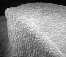
Photo used with
permission from K. Tosney
_________________________________________________________________________________________________
The Pierson’s Puppeteer
The Pierson’s Puppeteer has a morphology different from
that of a standard vertebrate tetrapod. While the
forelimbs are the same as any given tetrapod, the
hindlimbs are fused together to form one powerful unit.
The fusion is similar to individuals who suffer from
sirenomelia. Individuals with sirenomelia are
characterized by a fused lower limb as well as
deformities in the urogenital region; the deformities in
the urogenital region accounts for the severe lethality
of the disease (Kjaer 2003). In the Pierson’s
Puppeteer, the formation of the large single leg is due
to a replacement of the paired umbilical arteries with a
single large artery arising high from within the
abdominal aorta. As a result, the caudal end of the
developing Puppeteer would be deprived of oxygen leading
to the deformed single leg (Tang 1991). The Pierson’s
Puppeteer, unlike humans with sirenomelia, does not
suffer from the lethal urogenital defects. This is
because at the 9th week of development (the
earliest detectable age for sirenomelia) the single
umbilical artery undergoes apoptosis and divides into
two arteries (Schiesser 2003). This allows increased
blood flow into the limb regions and prevention of
profound urogenital defects. However, because the
single limb had already formed FGF will be concentrated
into a single patch at the rear of the animal creating a
single powerful leg.
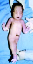
© KB Taori
(Click on Image to view Original Source)
Sources:
Altabef, Muriel and Cheryll Tickle. Initiation of dorso-ventral
axis during chick limb development. Mechanisms of
Development 116, 19-27. 2002.
Browder, Leon and Laurie Iten. Vertebrate Limb
Development. Dynamic Development. <http://www.ucalgary.ca/UofC/eduweb/virtualembryo/limb_dev.html>.
1998.
Kjaer, Klaus W. et al. Sirenomelia Sequence According
to the Distance Between the First Sacral Vertebra and
the Ilia. American Journal of Medical Genetics 120A:
503-508. 2003.
Schiesser, Monika et al. Sirenomelia, the mermaid
syndrome- detection in the first trimester. Prenat
Diagn, 23: 493-495. 2003.
Tang, Thomas et al. Limb Body-Wall Complex in
Association With Sirenomelia Sequence. American Journal
of Medical Genetics 41:21-25. 1991.
Taori, KB et al. Sirenomelia Sequence (Mermaid)- Report
of Three Cases. <http://www.ijri.org/articles/archives/2002-12-3/musculo_399.htm>.
Tosney, K and Pamela Raymond. Lecture Material,
November 28, 2005.
Xu, Xiaoling et al. Fibroblast growth factor receptor 2
(FGFR2)-mediated reciprocal regulation loop between FGF8
and FGF10 is essential for limb induction. Development,
125, 753-765. 1998.
 |