|
Eye Induction
Mouse From “Earth” Has Eyes Directly On Body!
In
our investigation into the development of the mouse from
the planet Earth, we had a special focus on the eyes,
which are so different from our Puppeteer eyes. The
eyes of a mouse are located on either side of its head,
which along with the eye and mouth also houses its
brain. The eyes are stuck in the head and cannot look
in opposite directions as if our eyes can, but instead
must always look in the same direction.
How Do Mouse Eyes Develop?
Placement and Development of the Eyes:
Diencephalon
and optic vesicle.

Before neurulation, the three transcription
factors Six3, Pax6, and Rx1 are all expressed at the
neural plate’s most anterior portion. According to
Gilbert, “This single domain will later split into the
bilateral regions that form the optic vesicles.” After
neurulation, this anterior portion of the neural
tube forms the mouse brain. The optic vesicle develops
from the portion of the brain called the diencephalon
(Gilbert). The optic vesicle reaches the ectoderm of
the head region of the chick, and through direct contact
with the ectoderm will induce the eye. Pax6, a
transcription factor, localizes in the optic vesicle
portion of the diencephalon and the ectoderm that will
form the lens. This confers competence onto the
epithelial ectoderm tissue so that it can form
the lens; it also confers specificity because only
the epithelial ectoderm will be able to form the lens
(Gilbert). The neural ectoderm thickens and causes the
epithelial ectoderm to thicken and form the lens placode.
The lens placode invaginates and then induces the optic
vesicle to form the optic cup. The outer layer of the
optic cup will eventually become the pigmented retina,
while the inner layer of the optic cup will become the
neural retina. The ganglion axons of the retina find
their paths to the portion of the brain called the
lateral geniculate nuclei with the aid of Eph receptors
and Ephrin ligands (Gilbert, Tosney). SHH, a
transcription factor, is responsible for midline
structures and is involved in the development of the eye
and nose. It separates the single eye field into two
bilateral fields (Gilbert). In the absence of SHH, only
one eye forms in the middle of the face, and the nose
forms dorsal to this eye; this condition is called
cyclopia (Gilbert, Tosney).
Hollow Lens Vesicle
The cells of the lens form from the equator and grow out
to form a hollow ball that is the lens. The lens cells
make crystallins after they mature, and new cells are
added from the equator as the lens grows (Tosney). The
crystallins will fill the empty space inside the lens
(Gilbert).
Cornea
The cornea is made of epithelial ectoderm that is
induced by the lens. These cells form a primary matrix
of collagen, which mesodermal mesenchymal cells use as a
substrate to form the corneal endothelium. The
endothelium in turn secretes a secondary matrix of
hyaluronic acid and fibrin. Neural crest cells then
migrate in and secrete hyaluronidase to degrade the
hyaluronic acid; the cells become immobilized. The
cornea is not transparent until the matrix becomes
dehydrated through the action of a sodium pump turned on
by thyroxine. Interocular pressure causes its
characteristic curve to develop.
_________________________________________________________________________________________________
Puppeteer Development
In the typical mouse that we studied, the eyes are only
a short distance from the brain. Our eyes, however, are
located further away from our brains. How do they grow
so far away?
Placement and Development of the Eyes
Puppeteer eyes use modified pathways that are not unlike
murine eye development. In a typical Puppeteer, the
neural tube diverges at the anterior pole of the embryo
as it begins to develop into the brain. This process
accounts for our two-neck phenotype. Prior to this,
however, the diencehpalon begins to from two optic
vesicles, one on each side of the br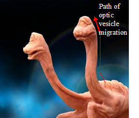 ain.
The formation of the vesicles stimulate the underlying,
medial neural tube cells to undergo rapid mitosis just
in those regions underlying the vesicles. Thus, the
neural tube diverges and then lengthens toward the
migrating anterior pole (to become the two heads). As
you may have noticed, the developing brain remains at
the base of the necks while the optic vesicles lead the
anterior migration of the two diverged neural tubes. ain.
The formation of the vesicles stimulate the underlying,
medial neural tube cells to undergo rapid mitosis just
in those regions underlying the vesicles. Thus, the
neural tube diverges and then lengthens toward the
migrating anterior pole (to become the two heads). As
you may have noticed, the developing brain remains at
the base of the necks while the optic vesicles lead the
anterior migration of the two diverged neural tubes.
This sequence of vesicle formation, divergence of the
neural tube, and migration of the newly formed anterior
tubes led by the optic vesicles assures that the
vesicles migrate to the correct location and confront
competent ectoderm for lens formation. From there,
development is the same as the eye of the mouse. This is
to say that once the optic vesicles confront competent
ectoderm they induce it to invaginate and form the lens
of the eye.
© Larry Niven, modified.
(Click on Image to view Original Source)
Sources:
Gilbert, Scott F. Developmental
Biology. 7th ed. Sunderland: Sinauer Associates, Inc.,
2003. Tosney, Kathryn W. “Eye development.” Biology 208:
Embryology, Ann Arbor. 8 Nov. 2005.
Tosney, Kathryn W. “Eye development.” Biology 208:
Embryology, Ann Arbor. 8 Nov. 2005.
|