|
Brain Growth & Induction
The mouse is a strange organism and
is not what most puppeteers would consider to be
normal. This organism has only one head which holds its
brain and both of its eyes. How does this strange and
simple animal’s brain develop? After months of research
we have pinpointed the major differences and now understand
why the mouse’s brain is located in its head instead of
its trunk.
As far as the development of the
brain, the mouse and puppeteer are almost identical. The
neural plate forms and begins to fold upwards. When the
two sides of the folding epidermis meet, the neural
tube is formed. After the neural tube closes there is a
build up of fluid in the anterior most section of the
neural tube which causes a large lumen to form. This
fluid pressure causes increased mitosis in the brain
region. The blockage of fluid is key in both mouse and
puppeteer brain development.
The
central nervous system (CNS) develops differentially
along both the anterior/posterior axis, and the
dorsal/ventral axis. The dorsal/ventral axis is
induced by a TGF-beta gradient supplied by the ectoderm
dorsal to the neural tube and Sonic Hedge Hog (SHH) is supplied by the notochord ventral to the neural tube.
Meanwhile, differential development along the anterior/posterior
axis is controlled by Hox gene expression.
The combination
of Hox gene expression
and ectodermal/notochord interactions cause the neural
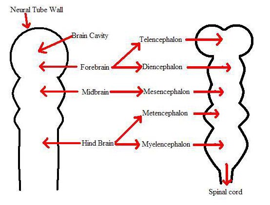
tube to development into several different layers and
regions. The neural tube differentiates into the forebrain,
mid-brain, and hind brain. These sections further
differentiate into the telencephalon, diencephalon,
mesencephalon, metencephalon, myelencephalon, and the
spinal cord, respectively.

Mouse Brain Development
Puppeteer Brain Development
The main difference between the
mouse and puppeteer brain formation is the placement of
the brain. The mouse’s brain forms at the anterior most
portion of the neural tube where as the puppeteer’s
brain is located at the base of the neck. In the mouse
one fluid block is necessary at the base of the brain.
In the puppeteer three fluid blocks are necessary. One
blockage is located at the base of the brain and the other
two blockages are located where the two anterior neural
tubes meet the diencephalon. After Henson's node
migrates, and the neural tube is formed, the
diencephalon then stimulates optic vesicle formation,
and thus mitosis of the underlying cells. This
induction by the diencephalon is not present in the
mouse and is the cause for only one head encasing the
brain.

Copp AJ, Greene NDE and Murdoch JN (2003). The genetic
basis of mammalian neurulation. Nature, 4, pp. 784-793.
Sources:
Gilbert, Scott F. Developmental Biology. 7th
ed. Sunderland: Sinauer Associates, Inc., 2003. p.
364-368
 |