The Rhodamine B molecules were excited by light from a modelocked 100 fs Titanium:Sapphire laser operating at 800 nm with a repetition rate of 80 MHz. A grating before the fiber coupler corrected for linear group velocity dispersion effects in the fiber and ensured that the pulse width at the fiber output remained 100 fs. Typically, uncoated probes with apex diameters of 200 nm were used.
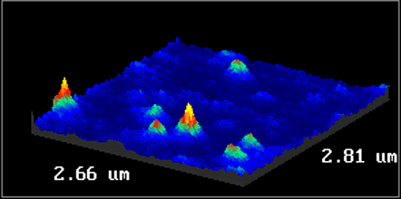
Figure 8 is an image of isolated single Rhodamine B molecules recorded by two-photon induced fluorescence using an average transmitted power of 150 mW. Scans using different excitation intensities confirmed the expected dependence of the fluorescence on the square of the intensity (data not shown).